How to Save a Tooth with Internal Root Resorption
What is Internal Root Resorption?
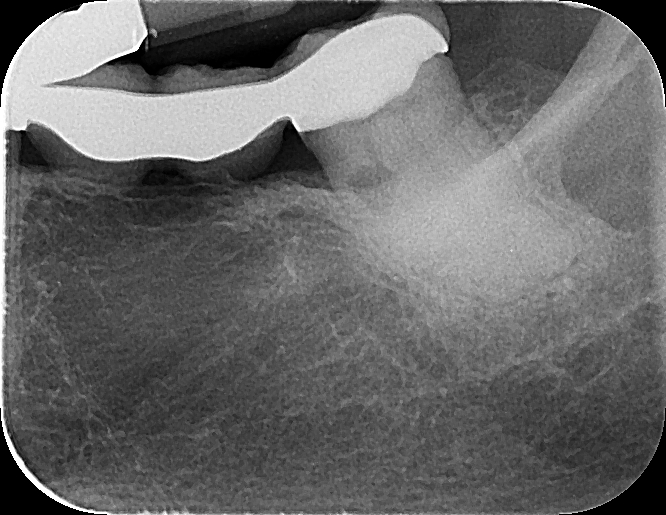
Internal Root Resorption is described as the loss of tooth structure inside the tooth, often caused by dental trauma or dental infection. If left untreated, resorption may continue to break down dentin and cementum until the tooth breaks or is no longer usable. Below is an example of how internal root resorption appears on on a diagnostic x-ray, taken from a recent case involving tooth #17.
The dark round spot on the left side of the tooth is a sign of internal resorption
How do you treat Internal Root Resorption?
Resorption of bone and teeth in the human body is caused by osteoclasts, which are naturally occurring bone cells that progressively break down bone tissue. These osteoclasts will continue to break down tooth structure as long as they are continually “fed” by surrounding tissue and blood vessels. In order to halt further bone degradation, root canal therapy is performed and the pulp is removed in order to cut off the osteoclasts’ “food supply”. Teeth suffering from internal resorption can be saved this way if treated in the early stages.
Did you know?
Osteoclasts are responsible for baby teeth to loosen and naturally fall out
How CBCT Technology Is Used
CBCT (Cone Beam Computed Tomography) is a type of CT scan that allows the endodontist to produce a 3D x-ray of the tooth. Cone Beam scanner emits much lower radiation compared to other CT scanners that are typically used in other branches in medicine. Below, you can see how the endodontist manipulates the scan to view the tooth in 3D and inspect the extent of the resorption in great detail. From the CBCT scan, the endodontist was able to identify the stage of resorption and determine the tooth was savable with root canal treatment.
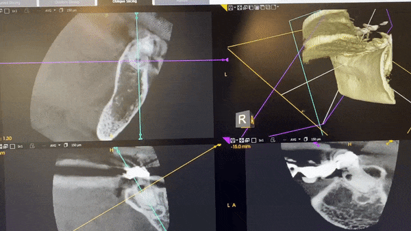
Viewing resorption of tooth in 3D with CBCT scan
Treatment Plan with Root Canal Therapy
The patient in this case was referred to RootVision Endo and was motivated to save the tooth and the existing bridge that was anchored by tooth #17. The root canal was completed by carefully creating a small access through the crown of the bridge so it can be preserved. Without the assistance of CBCT technology, the referring dentist may have recommended extraction based on the diagnostic 2D x-ray.
Diagnostic x-ray before root canal treatment 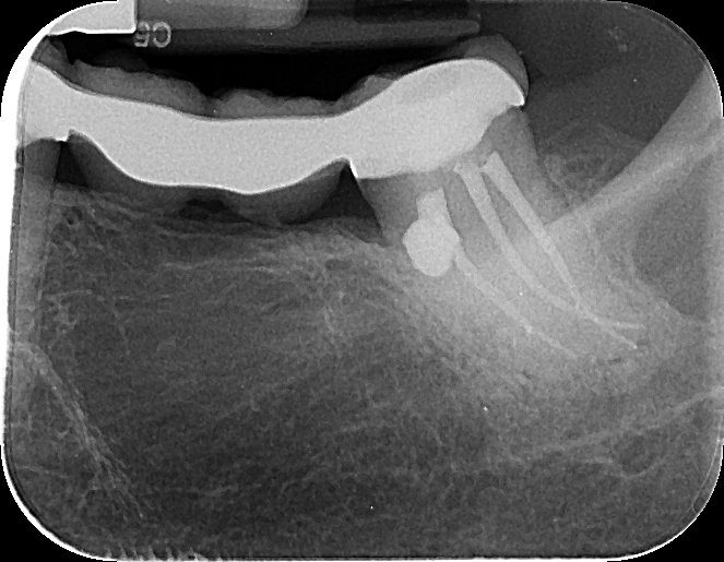
Post-op x-ray after root canal treatment